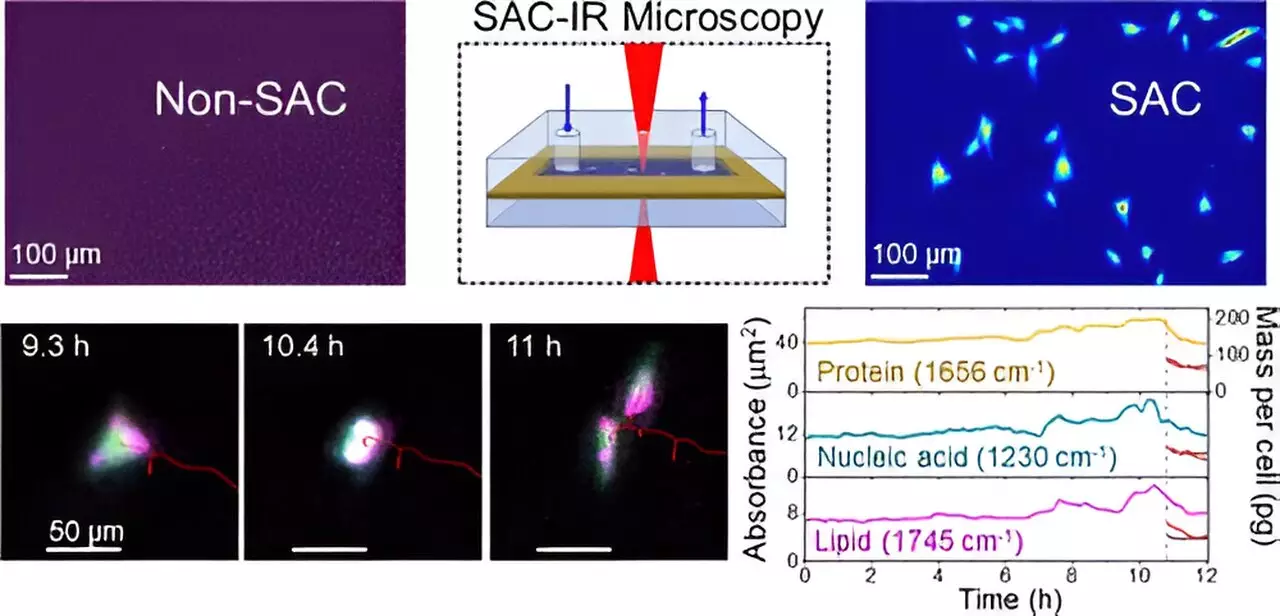
Biotechnology is at the forefront of medical advancements, offering groundbreaking drug therapies and cell-based treatments that promise to alter the trajectory of patient care. To harness the full potential of these innovations, researchers have focused their efforts on improving the ways we observe biomolecules within living cells. A recent development spearheaded by the National Institute of Standards and Technology (NIST) has introduced a revolutionary method using infrared (IR) light for imaging biomolecules, overcoming significant challenges posed by water’s interference in standard imaging techniques.
The imaging of biomolecules in their natural state has long been plagued by the presence of water, which is abundant among intracellular environments. Water strongly absorbs infrared radiation, making it difficult to detect other biomolecules that are of greater interest to researchers, such as proteins, lipids, and nucleic acids. In essence, the interference from water acts like a heavy fog, obscuring the finer details of cellular components. NIST’s researchers aimed to overcome this obstacle, allowing for clearer insights into the molecular composition and behaviors within living cells.
Introducing the Solvent Absorption Compensation Method
NIST chemist Young Jong Lee led the charge with the development of a new technique known as solvent absorption compensation (SAC). This method tailors the use of infrared light to selectively diminish the masking effects of water in IR measurements. By employing a specially designed optical system, SAC can isolate the absorption spectra of critical biomolecules, unveiling their presence amidst the heavy background noise created by water. This innovation transforms the way scientists can study cellular dynamics, shedding light on previously hidden interactions and functions of biomolecules.
The SAC method employs a hand-built IR laser microscope that excels in capturing detailed images of fibroblast cells—cells that play a crucial role in connective tissue formation—over a continuous observation period of twelve hours. This timeframe, while lengthy to the untrained eye, is considerably shorter than existing methods that depend heavily on extensive beam times at large synchrotron facilities. This efficiency not only accelerates the research pace but also enhances the accessibility of advanced biomolecular imaging.
A standout characteristic of the SAC-IR method is that it operates in a label-free manner. Traditional techniques frequently rely on dyes or fluorescent markers to visualize biomolecules, a practice that can potentially harm cellular integrity and reduce the accuracy of results. With SAC-IR, researchers can achieve absolute quantification of biomolecules—enabling them to measure proteins, nucleic acids, lipids, and carbohydrates without interference from extrinsic agents. This capability lays the groundwork for standardized analytical methods in various disciplines, including biology and medicine.
The implications of this technology are vast. For instance, in the field of cancer therapy, it is crucial to ensure the safety and effectiveness of modified immune cells prior to their reintegration into patients. New insights from the SAC-IR method can significantly enrich our understanding of the biomolecular changes that occur during this modification, ultimately providing a more accurate assessment of cellular health.
Expanding the Horizons of Drug Discovery
The transformative impact of NIST’s imaging technique extends to drug discovery and safety assessments. With the ability to measure the absolute concentrations of vital biomolecules within individual cells, researchers can employ this method to gauge the potency of new drug candidates and understand cellular responses to various treatments. Whether it’s evaluating a new pharmaceutical’s effectiveness or exploring drug interactions across diverse cell types, the SAC-IR method stands to offer an unprecedented level of clarity in drug screening processes.
As researchers continue to refine this technique, expectations are high for its potential to accurately measure other key biomolecules, including DNA and RNA. The relevance of this advancement goes beyond mere imaging; it opens new avenues in cellular biology. With improved measurements, scientists can begin to address fundamental questions about cell viability, providing insights into the mechanisms of life and death at the cellular level.
The Path Ahead: Broader Applications and Future Research
Looking forward, the continued development of the SAC-IR technology hints at a future where complex biological processes can be studied in detail, potentially revolutionizing how we perceive live cell interactions. Further research could help optimize preservation techniques for cells during freezing and thawing processes, which remain a considerable challenge in biotechnology.
The advancements made by NIST researchers signal a significant leap in our ability to observe and understand biomolecules in living systems. As this technology evolves, it promises to enhance biomanufacturing, improve drug development processes, and contribute to breakthrough therapies that leverage our understanding of cellular mechanics. This is not merely a step forward in imaging; it’s a leap towards realizing the full potential of biotechnology in harnessing life-saving innovations.


Leave a Reply