Understanding the intricate workings of a cell has long been a formidable challenge for scientists, primarily due to the limitations of traditional microscopy. Traditionally, microscopes could only resolve images down to approximately 200 nanometers, which is far too large to visualize many of the fundamental structures within cells. However, a collaborative innovation between researchers at the Universities of Göttingen and Oxford, along with the University Medical Center Göttingen (UMG), has marked a pivotal advancement in this field. Their groundbreaking work has led to the development of a microscope capable of achieving resolutions better than five nanometers, pushing the boundaries of what is visible.
Understanding Nanoscopic Dimensions
To grasp just how remarkable this advancement is, consider the scale of a nanometer. A single nanometer is one-billionth of a meter, which can be metaphorically compared to the diameter of a hazelnut relative to that of the Earth. This colossal difference in scale illustrates why traditional microscopy has struggled to provide detailed images of intracellular structures. Structures such as cytoskeletal filaments, which may measure only seven nanometers across, and synaptic clefts ranging from 10 to 50 nanometers pose official challenges to conventional methods, leading to fragmented and imprecise representations of cellular anatomy.
The newly developed microscope promises to revolutionize our understanding of these critical structures. With its sophisticated capabilities, it can provide comprehensive images of cellular components that were previously overlooked or merely inferred.
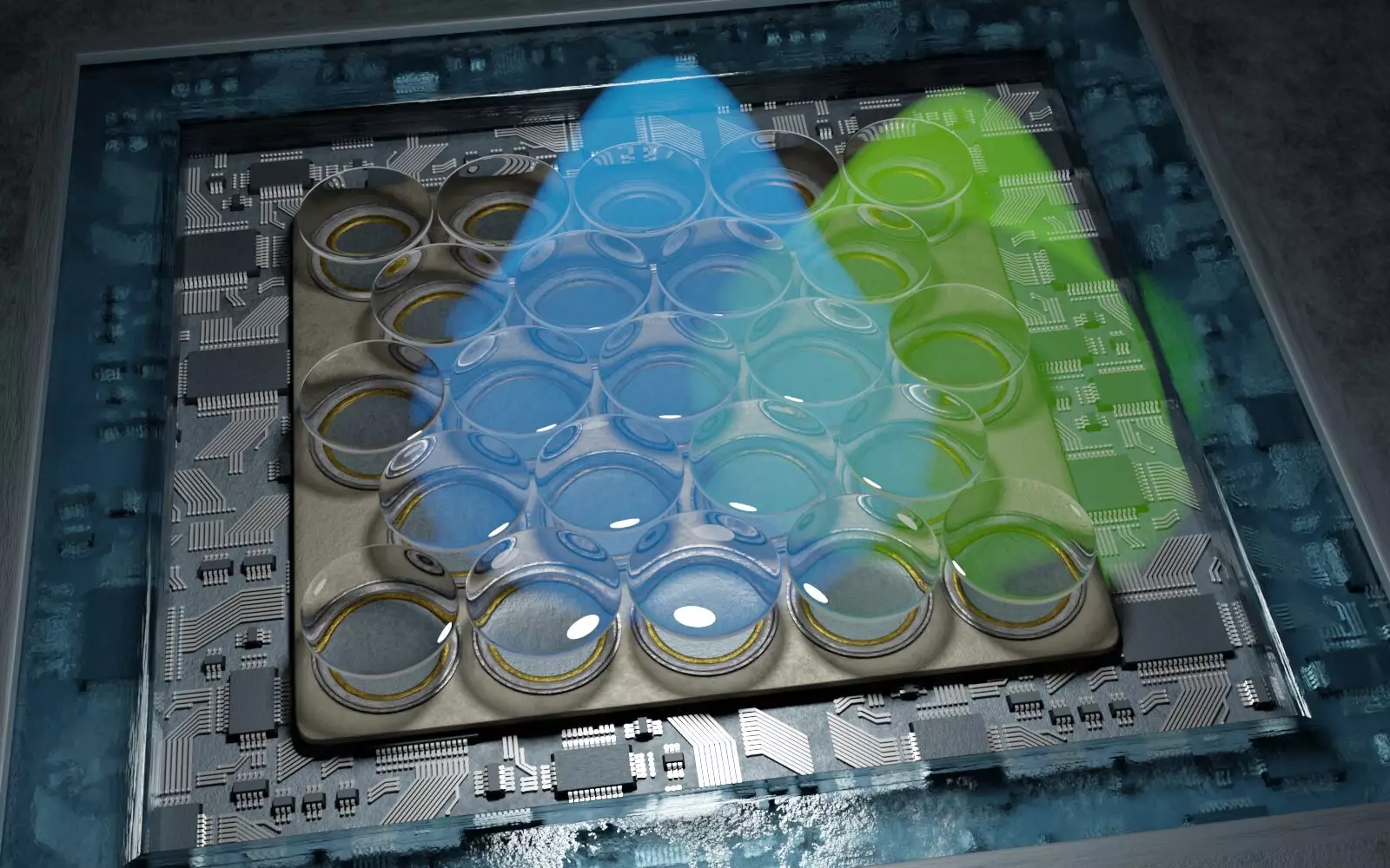
The technology behind this innovative microscope is rooted in a technique known as single-molecule localization microscopy (SMLM). This method involves the precise control of individual fluorescent molecules within a sample, switching them on and off in a systematic manner to accurately ascertain their locations. By gathering data on the positions of these molecules, researchers can rebuild a detailed model of the entire cellular structure.
Traditionally, SMLM achieved resolutions in the range of 10 to 20 nanometers. However, through the incorporation of a highly sensitive detector and specialized data analysis techniques, the team at Göttingen has managed to double this resolution. This newfound precision allows scientists to explore the minutiae of cellular interactions, including the organization of proteins in the synaptic junctions between nerve cells.
One of the significant advantages of this new microscopy technique is its cost-effectiveness and ease of use compared to other high-resolution microscopy methods. By offering a more accessible approach to high-resolution cellular imaging, it opens the door for a broader range of researchers in various biological fields, ultimately accelerating scientific discovery.
Professor Jörg Enderlein, a leading figure in this research, highlights that not only does this technology set a new benchmark in high-resolution microscopy, but it also democratizes access to these advanced imaging capabilities. In line with this pursuit of accessibility, the team has developed an open-source software package to facilitate data processing, further ensuring that researchers across disciplines can benefit from this technological leap.
The Implications for Future Research
The implications of this technology are profound. By providing a clearer understanding of cellular structures and their functions, researchers can delve deeper into the biological processes that underpin health and disease. This could lead to new therapeutic strategies and modalities in fields ranging from neuroscience to cancer research.
The advancements in microscopy achieved by the collaborative efforts of the Göttingen and Oxford teams mark a significant milestone in our quest to understand life at the cellular level. This innovation not only exemplifies the power of interdisciplinary research but also underscores the importance of accessibility in scientific advancement. As we stand on the brink of a new era in cellular imaging, the potential to uncover previously hidden biological intricacies is limitless.
Leave a Reply