Recent advancements in microscopy, particularly led by an esteemed team from Trinity College Dublin, are set to redefine multiple scientific fields, spanning from materials science to biomedicine. Their pioneering imaging technique employs advanced microscopy technologies to facilitate rapid imaging while dramatically lowering the radiation exposure to sensitive materials. This breakthrough stands as a testament to the continuous evolution of imaging technology, marking a significant stride toward achieving high-quality images without compromising the integrity of delicate biological tissues.
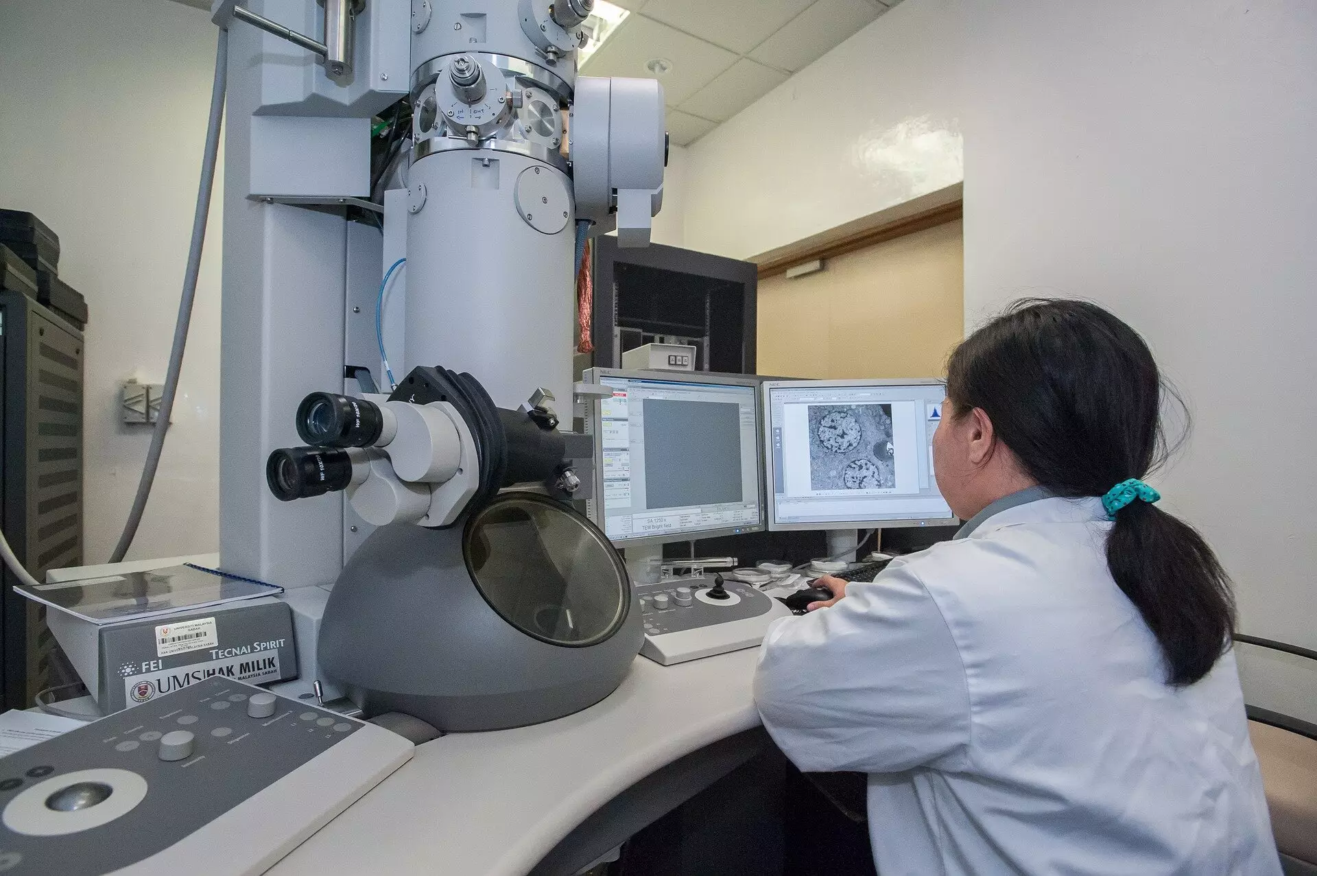
Conventional scanning transmission electron microscopy (STEM) techniques rely heavily on a focused beam of electrons that incrementally scans through a specimen, accumulating data pixel by pixel. This method, although reliable, typically adheres to a predefined dwell time at each pixel, which can translate into excessive radiation exposure. Much like a camera that uses constant exposure, STEMs subject all regions of the sample to the same level of intensity, risking damage—especially in cases involving sensitive biological specimens.
In contrast, the innovative approach introduced by this international research team advocates for a groundbreaking shift in imaging mechanics. Instead of persisting with a fixed time interval for signal accumulation, the new method pivots towards an event-based detection framework. Here, the image building process prioritizes the timing of the first detected electron at each pixel. This revolutionary change signifies a deep theoretical insight: the initial electron provides profound informational value, while subsequent detections yield rapidly diminishing returns. Consequently, this prompts the ability to minimize radiation exposure without sacrificing the quality of the image.
Cutting-Edge Technology and Its Implications
What further enhances the significance of this development is the accompanying patented technology, known as Tempo STEM. This reflects a synthesis of two advanced methods and incorporates an innovative “beam blanker” capable of toggling the electron beam with nanosecond precision in response to real-time imaging requirements. Such capability permits researchers to ‘shutter’ the beam at the optimal moment, hence obviating unnecessary radiation exposure and ultimately extending the viability of sensitive samples. This novel measure distinctively addresses the paradox inherent in traditional microscopy—wherein generating high-quality images conversely escalates the risk of irreparably damaging the samples involved.
Dr. Lewys Jones, who is an authority in the field and a pivotal figure in the team’s research, articulates this advancement as a “real leap” in understanding and capability. The technique not only reduces radiation doses but also streamlines the imaging process. The implications of this method, particularly in studies that involve fragile biomedical specimens, could be transformative, potentially leading to clearer and more accurate findings. It’s a chance for researchers to glean insights that were previously obscured due to the limitations of traditional techniques.
The Broader Impact on Biomedical Research
The repercussions of this new imaging technique extend beyond mere convenience or operational efficacy. In the realm of biomedical research, where the stakes of utilizing electron microscopy are particularly high, this approach could empower scientists with the ability to explore intricate cellular structures with minimal risk to the sample integrity. Often, traditional methods would lead to images that were either defective or misleading, rendering them unsuitable for crucial analyses.
Dr. Jon Peters, a key contributor to the work, emphasizes the critical nature of addressing electron-induced damage, especially at high speeds that could escalate the risk of harming biological samples. The discovery of a more efficient way to obtain these images opens new avenues for understanding complex cellular interactions and disease mechanisms at unprecedented resolutions. This could enhance diagnostics, inform therapeutic interventions, and elevate the overall quality of research outputs.
The merger of theoretical insight with practical application in this new imaging technique heralds a promising future for both scientific inquiry and clinical advancements. As the field of microscopy stands at the cusp of a technological renaissance, this innovative approach by Trinity College Dublin’s team exemplifies the power of creativity in scientific exploration, paving the way for a brighter, more accurate, and less destructive path forward.
Leave a Reply