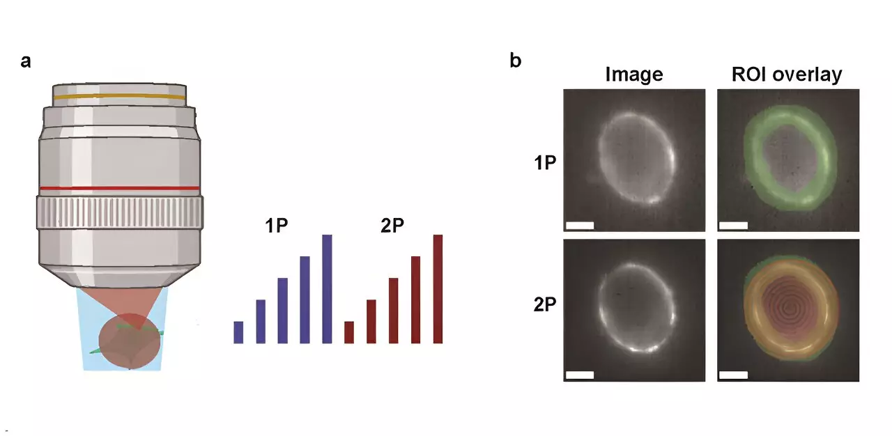
Understanding neural circuits is pivotal for comprehending how the brain processes information and facilitates communication between neurons. Recent advancements in imaging technology have enabled scientists to visualize neuronal electrical activity using genetically encoded voltage indicators (GEVIs). These GEVIs offer a unique lens through which researchers can observe the dynamic interactions occurring within the brain. However, the choice between two prominent imaging techniques—one-photon (1P) and two-photon (2P)—raises critical questions concerning their efficacy in real-world applications.
A landmark study from Harvard University has ignited renewed interest in the comparative effectiveness of 1P and 2P voltage imaging. Published in the journal Neurophotonics, this research delves into the optical and biophysical limitations of each method. The researchers undertook an exhaustive comparative analysis of the two techniques, focusing on the attributes such as brightness and voltage sensitivity inherent to widely adopted GEVIs under distinct illumination conditions.
Their key objective was to explore how fluorescence intensity diminishes with depth within mouse brain tissue, a concerning issue that persists in in vivo imaging experiments. Moreover, the team designed a predictive model to estimate the number of observable cells based on crucial parameters like reporter properties, imaging settings, and desired signal-to-noise ratio (SNR). Such a model could help streamline experimental designs in neural imaging efforts.
One of the study’s most significant revelations was the stark contrast in power requirements between the two imaging modalities. The researchers found that achieving a comparable photon count in 2P imaging necessitated around 10,000 times more illumination power per cell than 1P systems. This substantial power demand presents a double-edged sword, as it can lead to issues related to tissue photodamage and inherent shot noise, ultimately complicating data collection.
For instance, when examining the JEDI-2P indicator in the context of mouse cortex imaging—targeting a SNR of 10, with a bandwidth of 1 kHz, and laser power constraints—2P imaging struggled to monitor more than a dozen neurons simultaneously at greater depths, specifically beyond 300 micrometers. Such limitations severely restrict the potential for comprehensive neural exploration in living tissues.
The study concludes by asserting that current GEVIs’ modest voltage sensitivity combined with stringent photon-count needs makes in vivo 2P voltage imaging a formidable undertaking. To truly advance the field, significant advancements in 2P GEVIs are necessary, or an exploration into novel imaging methodologies should be pursued.
Through the insights gleaned from this examination, researchers are now better equipped with a nuanced understanding of the trade-offs between 1P and 2P voltage imaging. This work sets the stage for future technological advancements that promise to unravel the complexities of neural activity, ultimately enhancing our grasp of cognitive functions and disorders. Sustained innovation in imaging tools is essential for propelling our knowledge forward in the intricate world of neural circuits.
Leave a Reply